Quetip Focus Technique How It Works Might Surprise You

How the Q-tip focus technique works
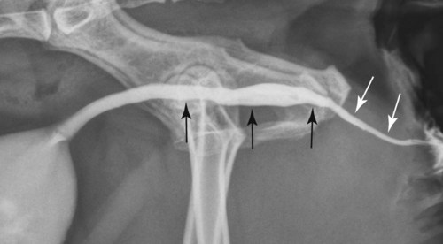
The Q-tip focus technique is a simple alignment method used in dental imaging to help position the X-ray beam correctly so it passes through tooth contacts cleanly and reduces overlap on bitewing images. In practice, a cotton swab acts as a visual guide for the angle of the beam, then the sensor holder is aligned to that same angle before the final image is taken.
The idea is not that the Q-tip itself does any imaging work; it is a reference marker. Clinicians use it to find the line that would travel straight through the contact points between teeth, then keep the X-ray tube head parallel to that line so the beam hits the target space instead of clipping tooth structure.
Why it is used
The main goal of the focus technique is to make bitewing radiographs more accurate and more readable by opening interproximal contacts. When contacts are overlapped, decay between teeth can be missed or become harder to judge, so a quick alignment aid can save retakes and improve diagnostic value.
This technique is especially helpful for students and early-career dental staff because it turns a hard-to-visualize beam angle into a physical cue that can be seen and matched in real time. The method is also practical because it uses a common, inexpensive object rather than specialized measuring tools.
How it works step by step
The process follows a three-part sequence: first, align the Q-tip with the correct contact angle; second, match the X-ray tube head or PID to that same angle; and third, place the sensor holder and expose the image without changing that relationship. In the source demonstration, the tube head and Q-tip are treated like parallel tracks, so the beam can be transferred safely from the visual guide to the final imaging setup.
- Find the contact line you want to open, usually by visually checking the premolar or molar contact area.
- Lay the Q-tip across the occlusal plane at the angle that would pass straight through the contact.
- Bring the PID or tube head into the same orientation, then remove the Q-tip once the angle has been transferred.
- Place the bite block or sensor holder while keeping the bar and tube head parallel, then take the exposure.
What makes it effective
The key advantage of the Q-tip method is that it externalizes geometry. Instead of asking a learner to imagine the beam path in three dimensions, it gives a visible line that can be compared directly with the PID position, reducing guesswork and helping the operator maintain the correct angulation.
It also reduces the risk of swinging the tube head into a slightly different angle after the contact line has already been identified. The guidance in the demonstration emphasizes keeping the assembly moving as a unit rather than swiveling the tube head independently, because small changes can bring the beam off target and create overlap.
Practical limitations
The technique is useful, but it is not foolproof. It still depends on accurate judgment of the contact line, patient cooperation, and correct placement of the sensor holder, and the demo notes that the bar and PID must stay parallel or the image geometry can fail.
It is also technique-sensitive in crowded mouths, where anatomy, limited opening, or positioning of the bite block can make alignment harder. In those cases, the Q-tip is only a guide, not a guarantee, and the operator still has to verify that the final beam path and receptor placement are consistent.
Why people search for it
Searches for the Q-tip technique usually come from dental students, assistants, and instructors looking for a quick way to understand bitewing angulation. The phrase can sound unusual, but the underlying principle is ordinary radiography: match the beam to the interproximal space so the image shows open contacts and usable detail.
"Think of the Q-tip as a stand-in for the beam path: find the line, match the PID, then keep everything parallel."
Common mistakes
A common error is lining up the Q-tip visually but then rotating the tube head afterward, which breaks the parallel relationship and ruins the transfer of angle. Another mistake is placing the bite block without checking that the bar still matches the PID, because the final receptor position must preserve the geometry established earlier.
Operators also sometimes forget that the Q-tip is a temporary guide. Once the angle has been identified and transferred, the guide should come out before exposure so it does not interfere with the patient's mouth or the sensor holder.
Who benefits most
Dental trainees benefit most because the method teaches beam geometry in a hands-on way. Experienced clinicians may also use similar visual alignment strategies when they want a quick, repeatable way to standardize bitewing placement across different patients.
For patients, the benefit is indirect but real: better-aligned images can mean fewer retakes, less chair time, and a lower chance of missing important interproximal findings. That makes the technique useful not just as a teaching trick, but as a workflow aid in routine radiography.
Illustrative workflow
| Step | What the operator does | Purpose |
|---|---|---|
| 1 | Identify the target contact area. | Define the beam path. |
| 2 | Place the Q-tip across the occlusal plane. | Visually mark the angle. |
| 3 | Align the PID with the same line. | Transfer the angle to the X-ray tube head. |
| 4 | Remove the Q-tip and position the bite block. | Prepare for exposure without changing geometry. |
| 5 | Take the image with parallel alignment preserved. | Improve contact opening and image clarity. |
Bottom line in practice
The focus technique works by turning beam alignment into something you can physically see and match before exposing the image. If the Q-tip line, the PID, and the sensor holder all stay aligned, the result is a cleaner bitewing with better-open contacts and fewer geometry errors.
Key concerns and solutions for Quetip Focus Technique How It Works Might Surprise You
Is the Q-tip focus technique used in all dental X-rays?
No, it is mainly a teaching and positioning aid for bitewing-style alignment problems, especially when interproximal contacts need to be opened clearly. It is not a universal replacement for standard radiographic positioning rules.
Does the Q-tip stay in the mouth during exposure?
No, the Q-tip is used temporarily to find and transfer the angle, then removed before the image is taken. The final exposure is made with the bite block and PID aligned, not with the cotton swab still in place.
Why do contacts overlap on dental X-rays?
Contacts overlap when the beam angle is off, so the X-ray passes across tooth surfaces instead of straight through the spaces between them. The Q-tip method helps the operator find that straight-through path more reliably.
What is the main teaching value of this technique?
Its main teaching value is that it converts an abstract geometric concept into a visible alignment exercise. That helps learners understand how beam angulation affects image quality much faster than theory alone.